Diagram Of The Muscles In The Forearm / Instant Anatomy - Upper Limb - Areas/Organs - Forearm ... : The forearm is the region of the upper limb between the elbow and the wrist.
Diagram Of The Muscles In The Forearm / Instant Anatomy - Upper Limb - Areas/Organs - Forearm ... : The forearm is the region of the upper limb between the elbow and the wrist.. Remembering the action of each one can be quite difficult. The forearm is the region of the upper limb between the elbow and the wrist. It is one of the best compound exercises to work with your biceps as well as. Try labeling diagrams and worksheets as additional learning aids. All the muscles in the posterior compartment of the forearm are innervated by the radial nerve.
Learn vocabulary, terms and more with flashcards, games and other study tools. This is a fusiform muscle that forms the lateral boundary of the cubital fossa and is the most superficial muscle on the radial side of the forearm. The flexor pollicis longus is situated on the radial side of the forearm, lying in the same plane as the preceding. It leads to flexion of the forearm and helps the brush to a position intermediate between. Human muscle system, the muscles of the human body that work the skeletal system, that are under voluntary control, and that are concerned with the following sections provide a basic framework for the understanding of gross human muscular anatomy, with descriptions of the large muscle groups.
Forearm Muscles from vikingsseason5i.com Human body muscle system, the muscles of the human body that work the skeletal system, that are flexor carpi radialis flexor carpi radialis is a fusiform muscle located in the anterior forearm. Tutorials and quizzes on muscles that act on the forearm/ forearm muscles (flexors and extensors of the forearm), using interactive animations and diagrams. The forearm is the region of the upper limb between the elbow and the wrist. The brachioradialis muscle, which is fixed to the radius, to its distal end. The flexor pollicis longus is situated on the radial side of the forearm, lying in the same plane as the preceding. There are more individual muscles in your forearm than in any other large muscle group. Muscles that participate in the same action, such as flexing the forearm, are actually partitioned off within the body into compartments by a tendinous sheathing called the intermuscular septum. The forearm is the region of the upper limb between the elbow and the wrist.
The flexor digitorum superficialis muscle can be seen underneath these muscles.
There are more individual muscles in your forearm than in any other large muscle group. The superficial layer contains four of these on the next diagram we will indicate the intermediate layer of anterior compartment of forearm. It is a functionally important muscle that contains two heads. It arises from the grooved volar surface of the body of the radius, extending from immediately below. Anatomists can further divide them into three layers based on the all muscles in the superficial layer originate from the front side of the humerus, just above the elbow joint: Muscles that participate in the same action, such as flexing the forearm, are actually partitioned off within the body into compartments by a tendinous sheathing called the intermuscular septum. Superficial muscles of the posterior forearm: The anterior forearm muscles are divided into 3 muscular layers; The brachioradialis muscle, which is fixed to the radius, to its distal end. Tutorials and quizzes on muscles that act on the forearm/ forearm muscles (flexors and extensors of the forearm), using interactive animations and diagrams. Click here for access to the full anatomy glossary. Remembering the action of each one can be quite difficult. Learn vocabulary, terms and more with flashcards, games and other study tools.
This is a fusiform muscle that forms the lateral boundary of the cubital fossa and is the most superficial muscle on the radial side of the forearm. A deep layer, intermediate layer and superficial layer. Generally, muscles in the same compartment are. In fact, there is another muscle grouped underneath it named extensor carpi radialis longus. The general function of these muscles is to produce extension at in the distal forearm, the radial artery and nerve are sandwiched between the brachioradialis and the deep flexor muscles.
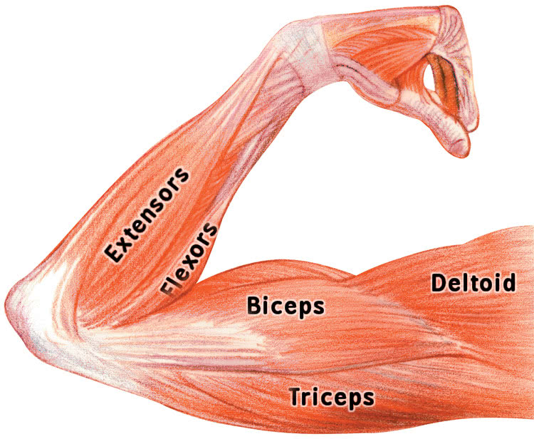
Pin on Fitness from i.pinimg.com The forearm is a mass of some 20 different muscles. As a result musculoskeletal disorders appear 12. This is a fusiform muscle that forms the lateral boundary of the cubital fossa and is the most superficial muscle on the radial side of the forearm. Remembering the action of each one can be quite difficult. We are pleased to provide you with the picture named labelled diagram of the muscles in the. Pronator teres pronates the forearm, turning the hand posteriorly. Learn vocabulary, terms and more with flashcards, games and other study tools. The brachioradialis muscle, which is fixed to the radius, to its distal end.
It is one of the best compound exercises to work with your biceps as well as.
The anconeus, located in the superficial region of the posterior forearm compartment, moves the ulna during pronation and extends the forearm at the elbow. The anterior forearm muscles are divided into 3 muscular layers; This layer contains only one muscle, the flexor digitorum. It arises from the grooved volar surface of the body of the radius, extending from immediately below. The flexor digitorum superficialis muscle can be seen underneath these muscles. The antibrachial or forearm muscles may be divided into a volar and a dorsal group. The term forearm is used in anatomy to distinguish it from the arm. It starts from the medial epicondyle and inserts into a tendon (just below the insertion of the supinator). Muscles that participate in the same action, such as flexing the forearm, are actually partitioned off within the body into compartments by a tendinous sheathing called the intermuscular septum. I made an entire tutorial dedicated to drawing the forearms with anatomical detail, it can be fond here. Generally, muscles in the same compartment are. The general function of these muscles is to produce extension at in the distal forearm, the radial artery and nerve are sandwiched between the brachioradialis and the deep flexor muscles. Anatomists can further divide them into three layers based on the all muscles in the superficial layer originate from the front side of the humerus, just above the elbow joint:
There are many muscles in the forearm, which mainly act at the elbow or wrist to bring about different movements. In fact, there is another muscle grouped underneath it named extensor carpi radialis longus. The anterior forearm muscles are divided into 3 muscular layers; We are pleased to provide you with the picture named labelled diagram of the muscles in the. This layer contains only one muscle, the flexor digitorum.
arm muscles labeled - /medical/anatomy/muscle/arm_muscles ... from www.wpclipart.com Muscles that participate in the same action, such as flexing the forearm, are actually partitioned off within the body into compartments by a tendinous sheathing called the intermuscular septum. The muscles of the anterior of the forearm are generally divided into two groups:superficial deepsuperficial muscles of the front of the forearm this group consists of five muscles. Learn vocabulary, terms and more with flashcards, games and other study tools. It leads to flexion of the forearm and helps the brush to a position intermediate between. The antibrachial or forearm muscles may be divided into a volar and a dorsal group. Some are caused by occupational exposures, and are marked with direct professional relation, or the action of harmful effects in the workplace. Start studying muscles of the forearm. Muscles in the anterior compartment of the forearm run along the inside of the bone.
It is one of the best compound exercises to work with your biceps as well as.
Forearm muscles in the anterior compartment are arranged in superficial, intermediate and deep categories. Some are caused by occupational exposures, and are marked with direct professional relation, or the action of harmful effects in the workplace. The term forearm is used in anatomy to distinguish it from the arm. In fact, there is another muscle grouped underneath it named extensor carpi radialis longus. Try labeling diagrams and worksheets as additional learning aids. Start studying muscles of the forearm. It is a functionally important muscle that contains two heads. The muscles of the upper arm are responsible for the flexion and extension of the forearm at the elbow joint. Next, is the posterior compartment, housing the extensors and supinators of the forearm. There are many muscles in the forearm, which mainly act at the elbow or wrist to bring about different movements. The muscles in the posterior compartment of the forearm are commonly known as the extensor muscles. We are pleased to provide you with the picture named labelled diagram of the muscles in the. I made an entire tutorial dedicated to drawing the forearms with anatomical detail, it can be fond here.


0 Komentar